Preliminary results

Rapid Critical Care Diagnostics Using Label-Free Imaging Cytometry

Nicholas Bratvold1, Ilakkiyan Jeyakumar1, Michelle Khoo1, Gregory Suematsu1, Olivia Chao2, Tal Shwartzman2, Carolyn Calfee2, Michael Matthay2, Natasha Spottiswoode2, Chaz Langelier1,2, and Paul M. Lebel1
1 Biohub  ·  2 University of California, San Francisco
These are unpublished, preliminary results. Subject to revision and not yet peer-reviewed.

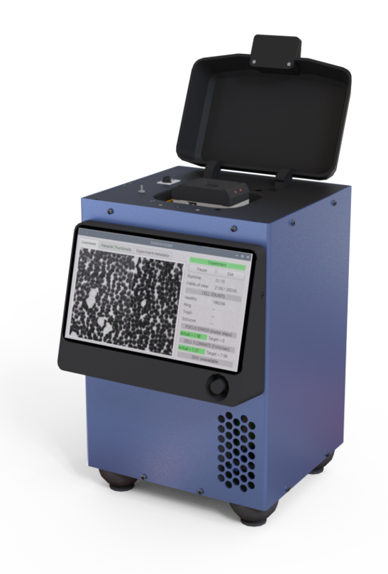
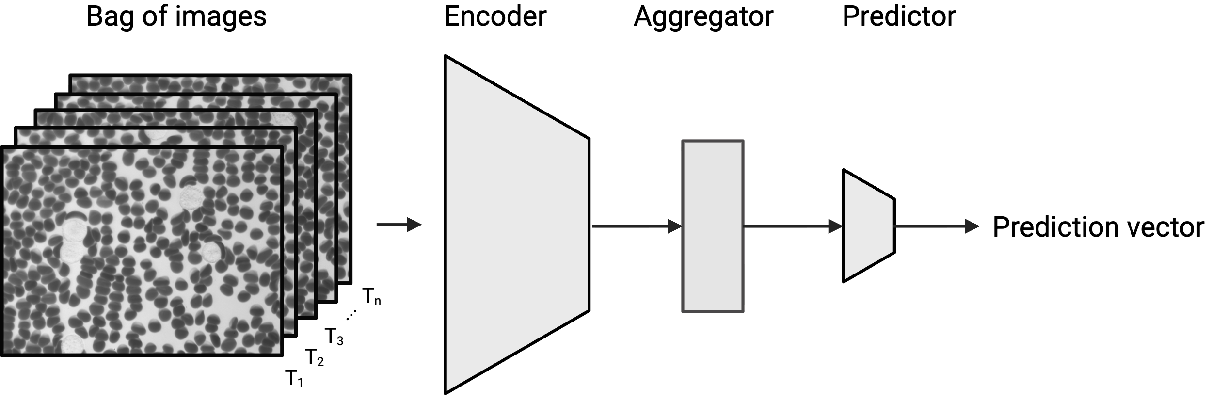
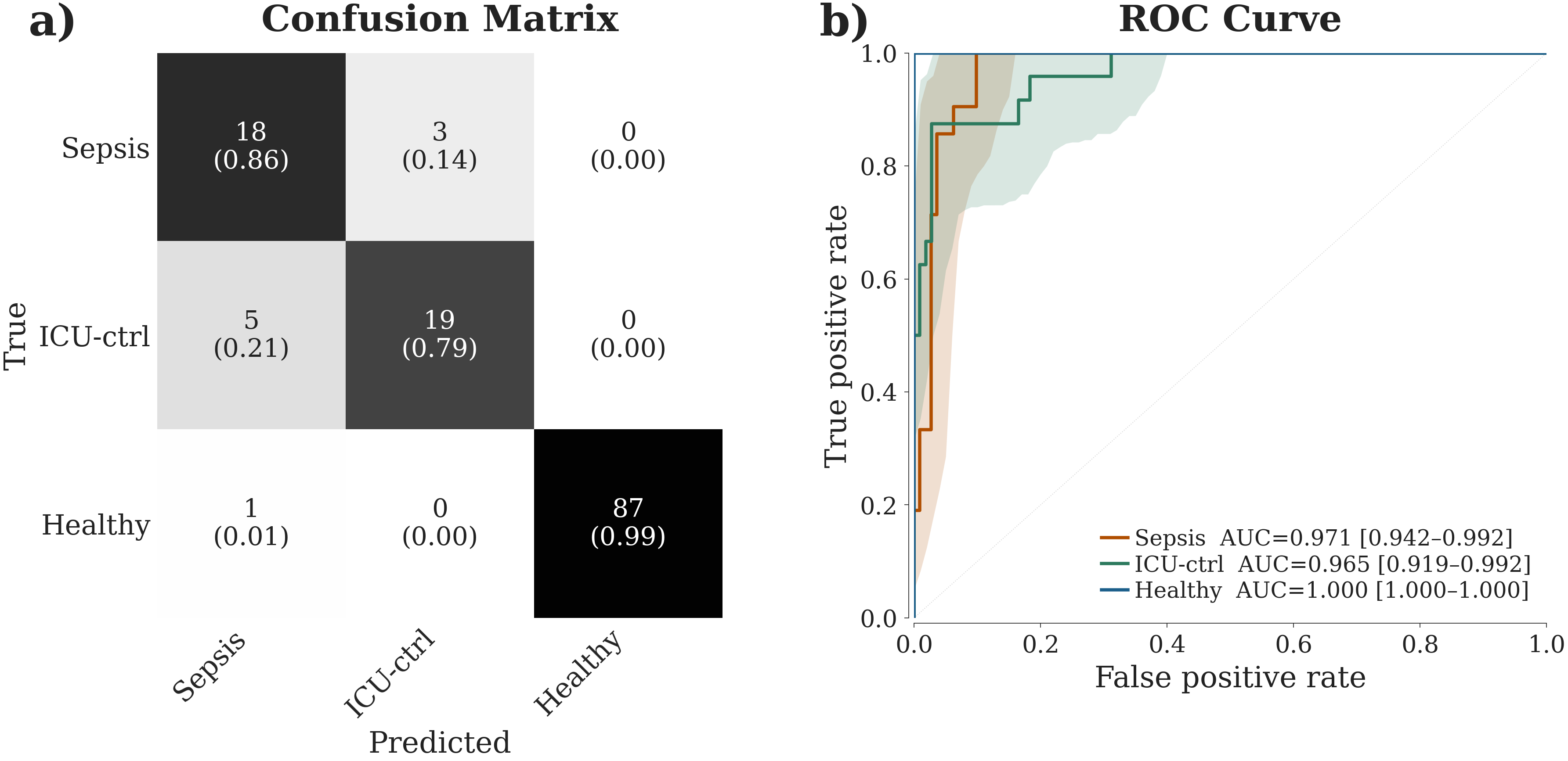
Remoscope analyzes millions of cells from fresh whole blood within minutes without fixation, staining, or any sample preparation. The remo-ID model learns to collect features across hundreds of images, that are predictive of the patient's physician-adjudicated disease state. We trained these models to distinguish sepsis-positive patients from other critically ill patients in the Early Acute Renal and Lung Injury cohort at the University of California San Francisco (EARLI), as well as from a pool of healthy blood donors. The model learns which features are relevant without being told what to look for. Across sepsis-positive patients, other critically ill patients, and healthy donors it reaches a macro F1 of 0.87, detecting sepsis at 86% sensitivity and 95% specificity.

Class activation

Where the model looks.

Sepsis

The red hue shows class-activation — where remo-ID looks when making a call. In these sepsis-positive cases, attention concentrates on abnormal and activated white blood cells. The images shown here are among the highest-confidence positive calls from a few patient samples.

P. falciparum (malaria)

We also trained this type of model to identify malaria (Plasmodium falciparum), which we previously studied using an object detection model called YOGO1. So far, with very little data, the remo-ID attention concentrates on infected red blood cells and overlaps very well with YOGO results. This strengthens our hypothesis that remo-ID is finding disease-predictive features and that extension of the method to many other diseases is possible.

Device & model

Device and Model

Remoscope imaging cytometer
Remoscope
remo-DB
remo-ID model architecture
remo-ID

Remoscope is a label-free imaging cytometer that cuts all sample preparation and user training out of the workflow. Fresh whole blood flows through a disposable ultrathin flow cell while millions of cells are imaged in minutes, with on-board compute running end-to-end automation and inference. Here we are able to accurately predict sepsis from just over a hundred images, which can be acquired in seconds.

Results

Quantitative Results

On a small dataset, we were able to train and validate remo-ID models that can distinguish sepsis from other critically ill patients and healthy donors with very high accuracy.

(a) Confusion matrix and (b) one-vs-rest ROC curves for the 3-class model
(a) Confusion matrix and (b) one-vs-rest ROC curves, 3-class model
Per-class performance — point estimate [95% CI]
ClassSupportF1Sens.Spec.PPVNPVLR+LR−
Sepsis21 0.800.67–0.91 0.860.69–1.00 0.950.90–0.98 0.750.57–0.91 0.970.94–1.00 16.008.37–49.70 0.150.00–0.32
ICU-ctrl24 0.830.69–0.93 0.790.62–0.95 0.970.94–1.00 0.860.70–1.00 0.950.91–0.99 28.7612.29–∞ 0.210.05–0.39
Healthy88 0.990.98–1.00 0.990.96–1.00 1.001.00–1.00 1.001.00–1.00 0.980.93–1.00 ∞–∞ 0.010.00–0.04
Macro133 0.870.79–0.95 0.880.80–0.95 0.970.95–0.99 0.870.79–0.94 0.970.95–0.99

All three classes are strongly separable, with one-vs-rest AUCs of 0.97 for sepsis [0.94–0.99], 0.97 for ICU-control [0.92–0.99], and 1.00 for healthy. Per-class F1 reaches 0.80 for sepsis, 0.83 for ICU-control, and 0.99 for healthy, for a macro F1 of 0.87. The sepsis class is detected at 86% sensitivity and 95% specificity, with a positive likelihood ratio of 16.

Morphology

remo-Map: Discovery of Cell Morphology Fingerprints

With remo-Map, we analyze millions of cells and send their images through a powerful embedding model to extract their features and separate them by cell type and state.2 As individuals, cells can be distinguished by type and state. As an ensemble, we can discover patient-level patterns occuring due to meaningful shifts in the distribution of cells that correlates well with their state of health.

Healthy
ICU-Sick
Sepsis

Here we plot millions of cells from the three classes (healthy, sick-icu, and sepsis), projected onto a UMAP via an unsupervised cell embedding. In sepsis, white blood cells shift darker and more granulated toward a state of activation.

Conclusion

Conclusion

The Remoscope, equipped with remo-Map and remo-ID, has allowed us to discover disease-predictive features in fresh blood samples that are highly amenable to point of care decision-making. This work is part of an ongoing research study, is subject to change as more results emerge, and will be submitted for peer review on completion along with all appropriate documentation and IRB approval for research on human subjects.

References

References

  1. Lebel, P. M. et al. Remoscope: a label-free imaging cytometer for malaria diagnostics. Trans. R. Soc. Trop. Med. Hyg. 119, 1100–1111 (2025). https://doi.org/10.1093/trstmh/traf070
  2. Bratvold, N. et al. Manuscript in preparation (2026).