Deep analysis of cell type and disease state.
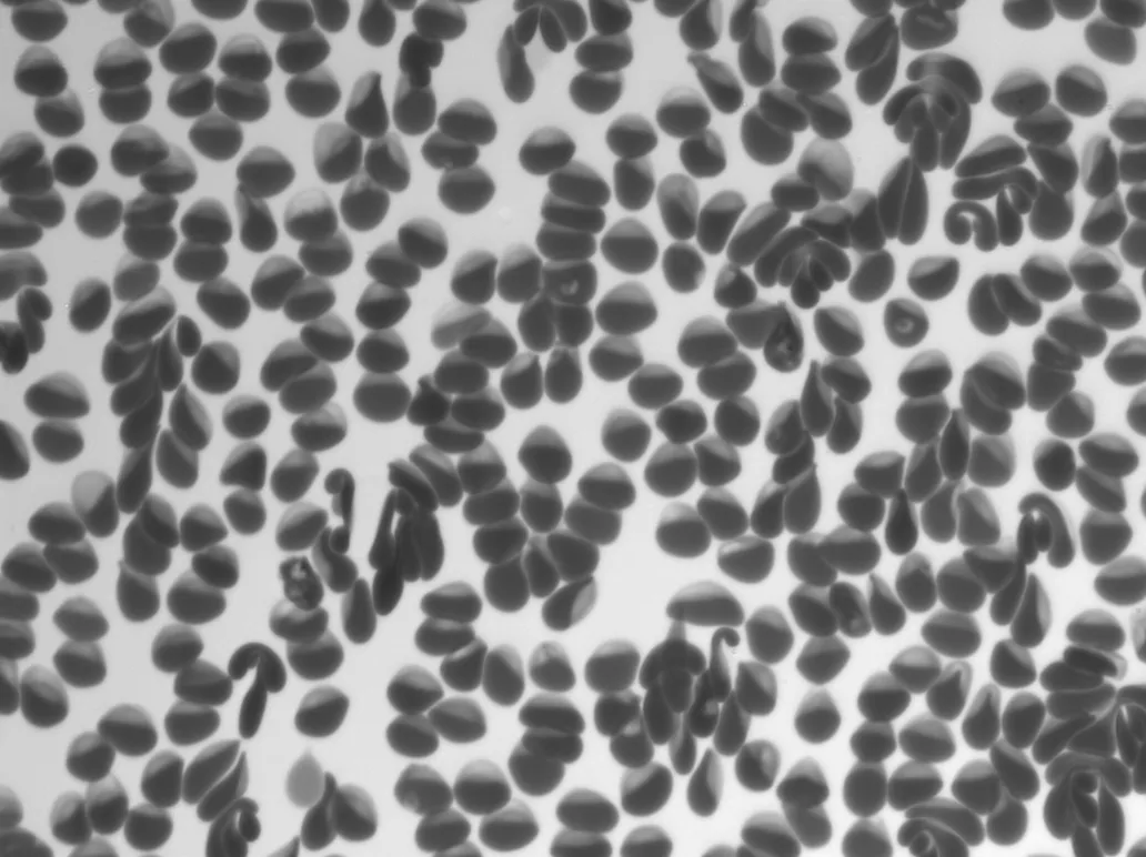
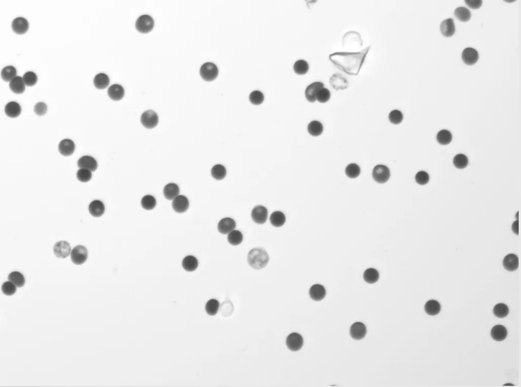
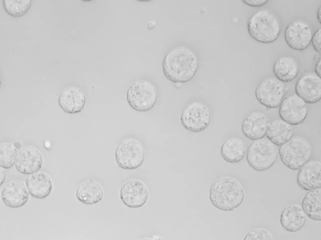
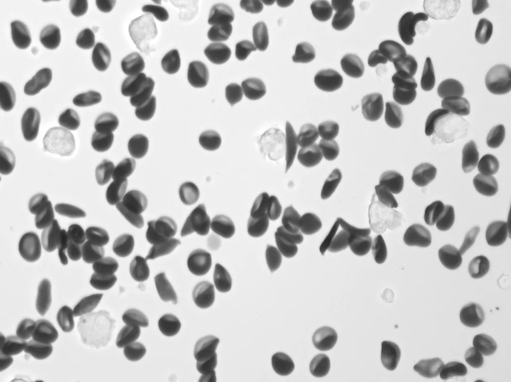
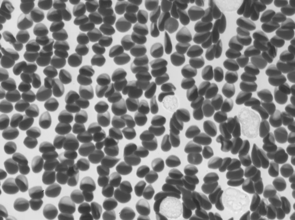
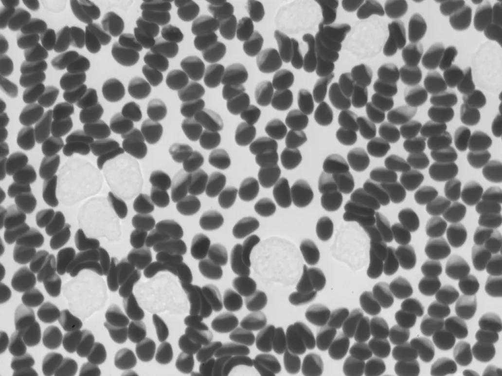
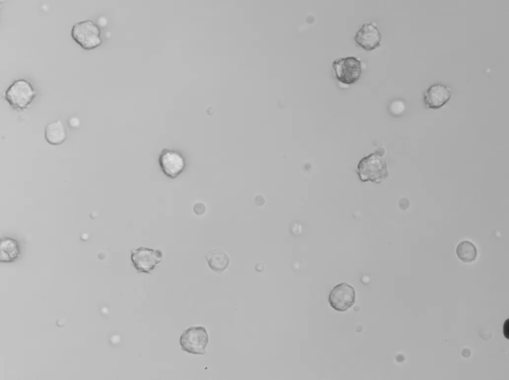
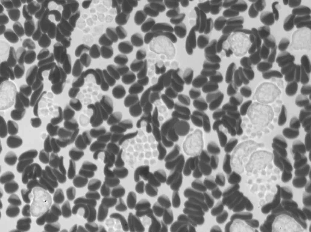
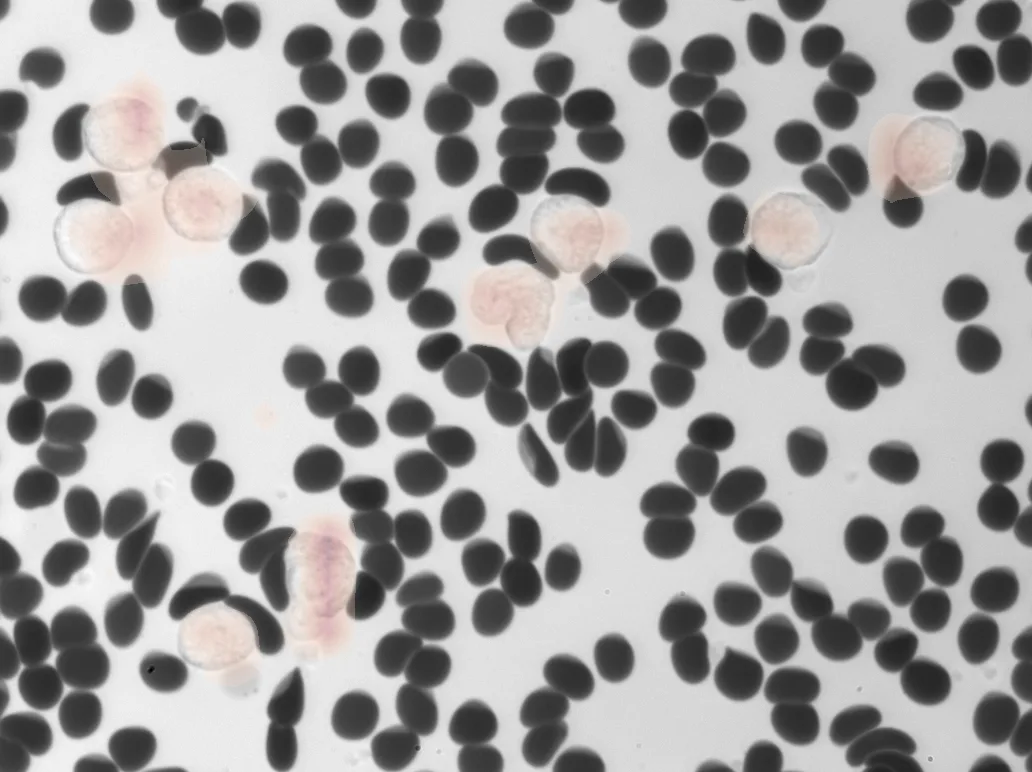
Remoscope images millions of cells from unprocessed fluid and analyzes them in real time, hands-free, with zero sample preparation.
-
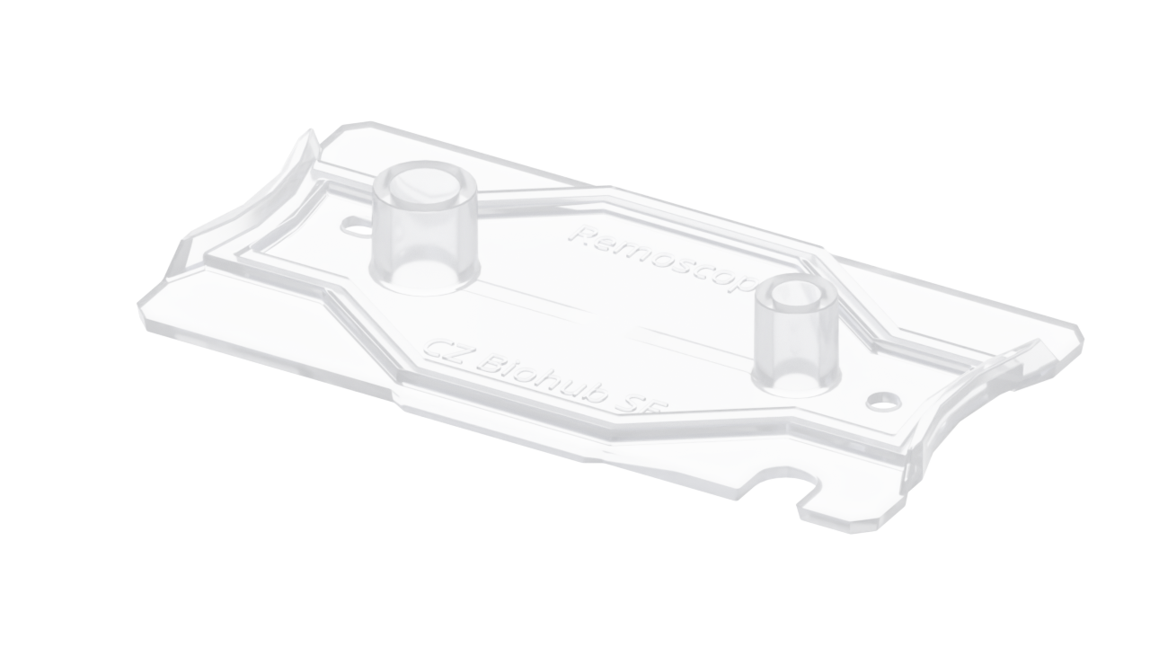 Consumable Ultrathin flow cell 10 µL, unprocessed, no preparation
 -
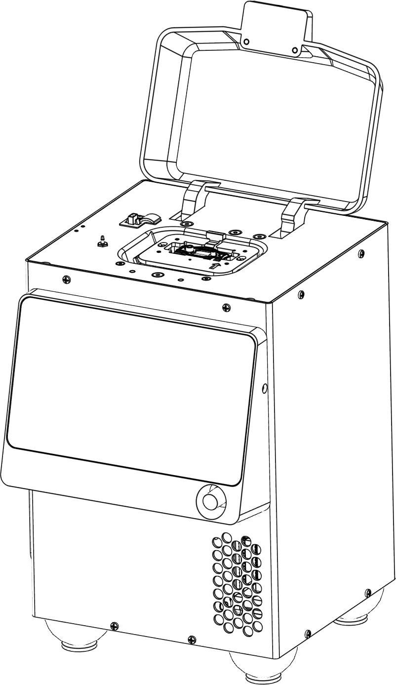
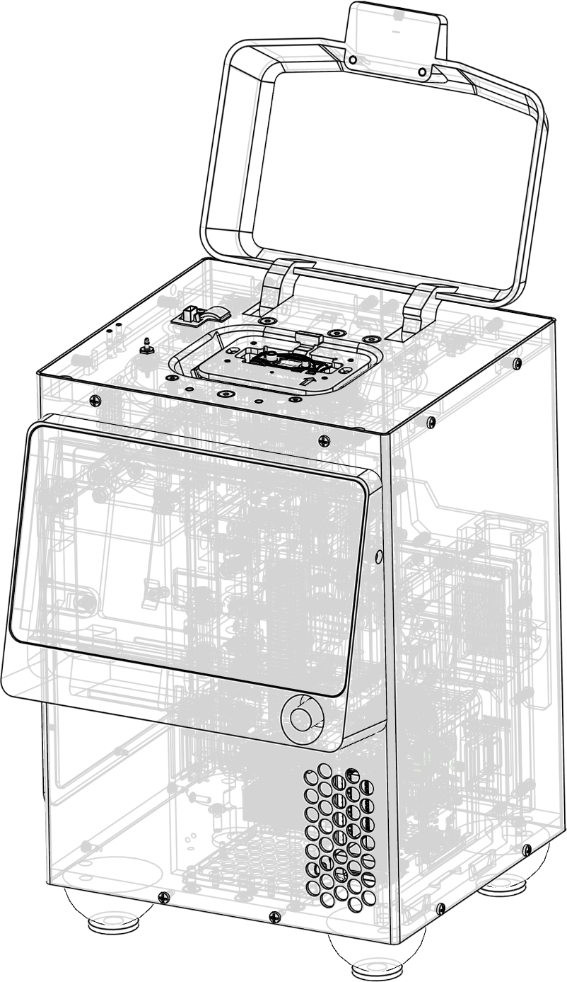
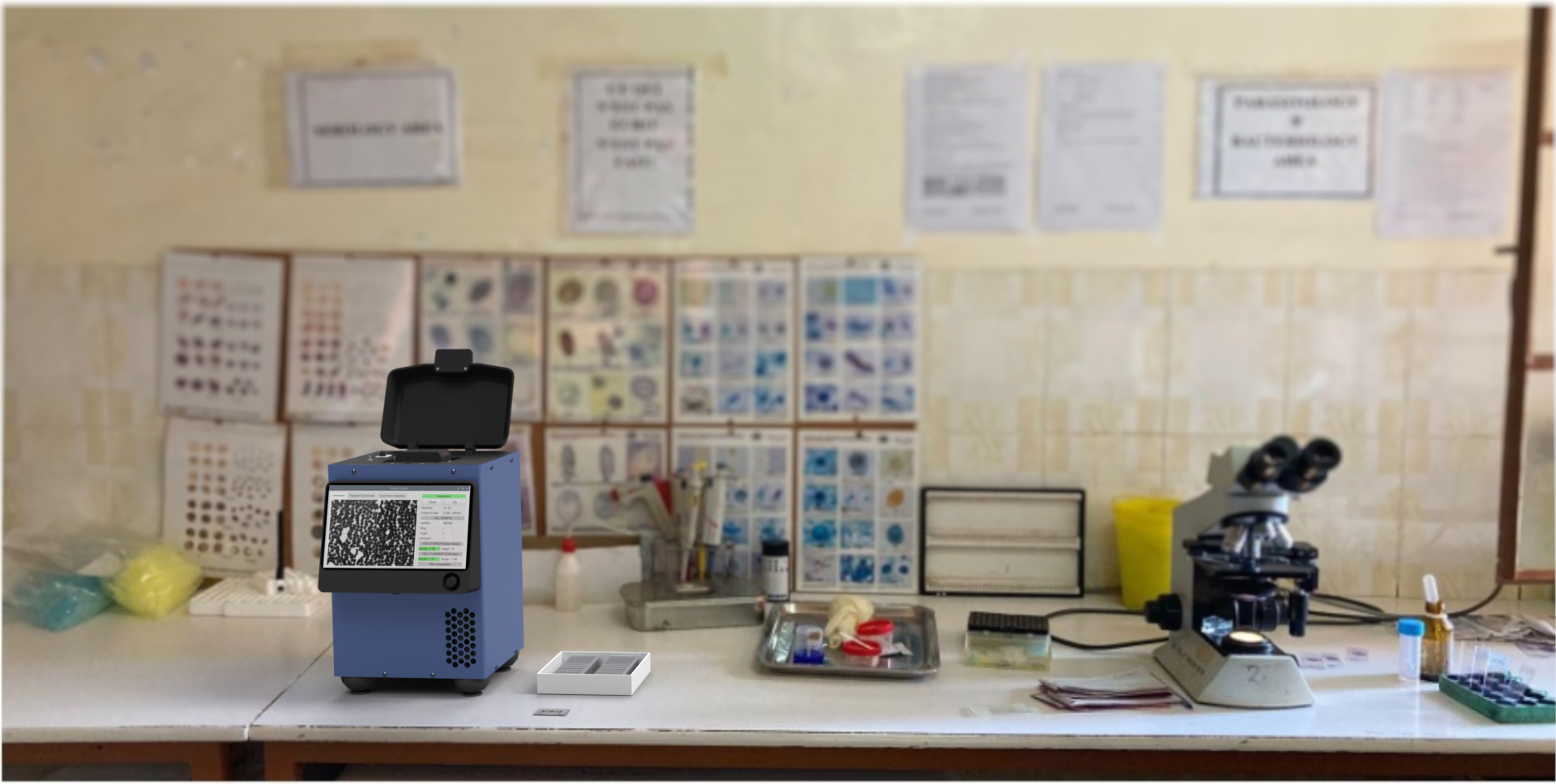
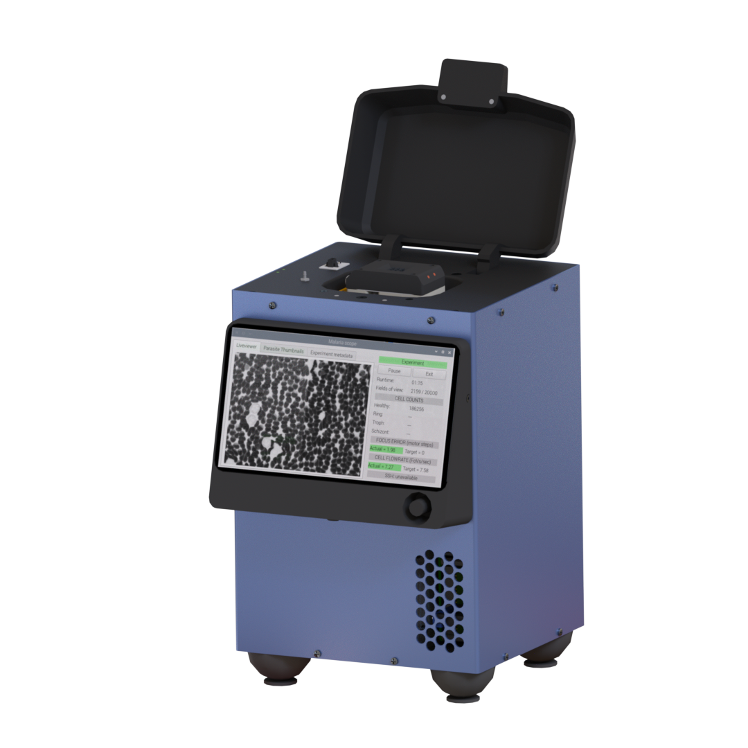 Hardware Remoscope A 200 × 300 mm benchtop device
 -
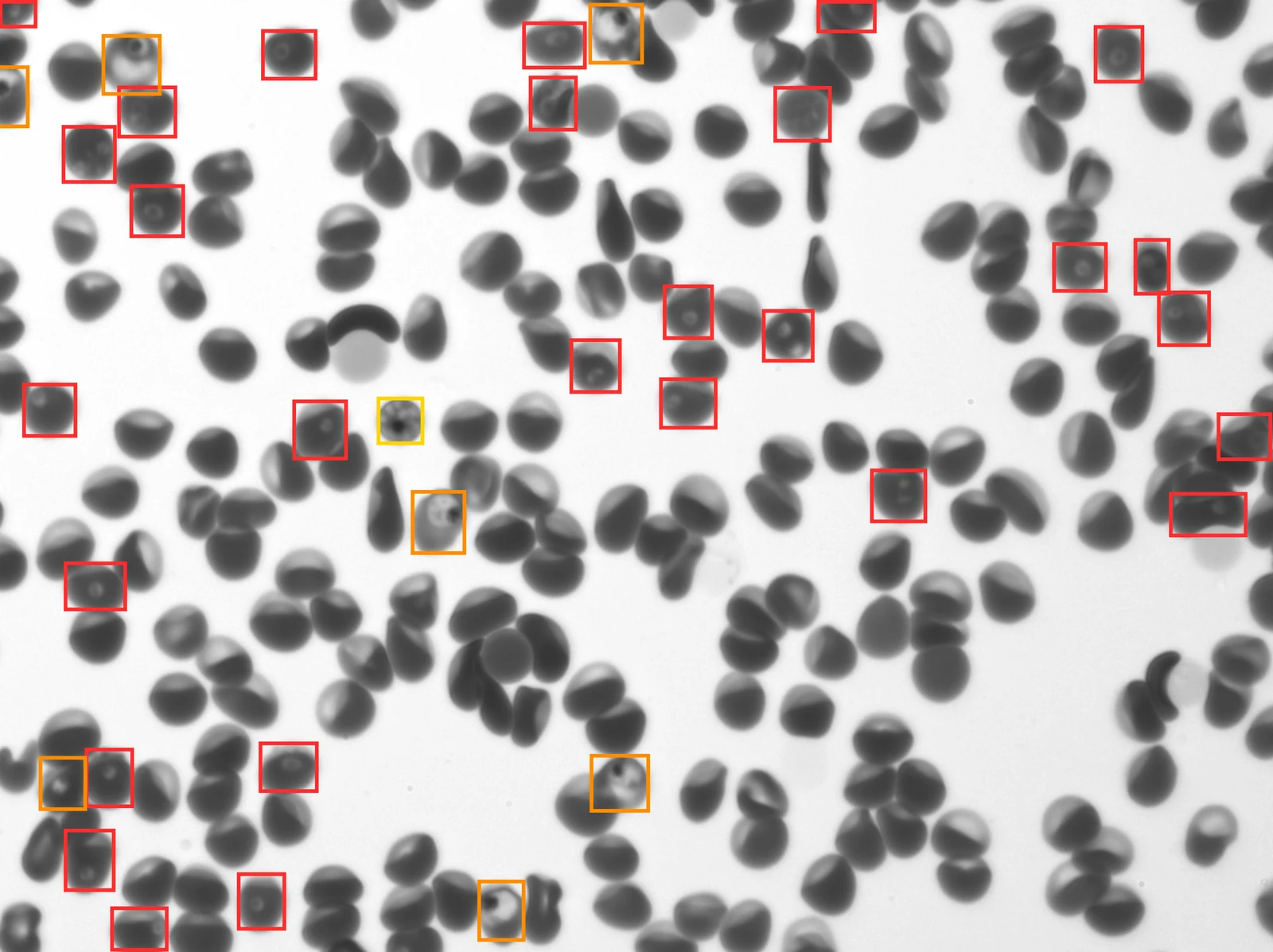
Data Every cell, imaged live 0 cells imaged (actual throughput!!)
-
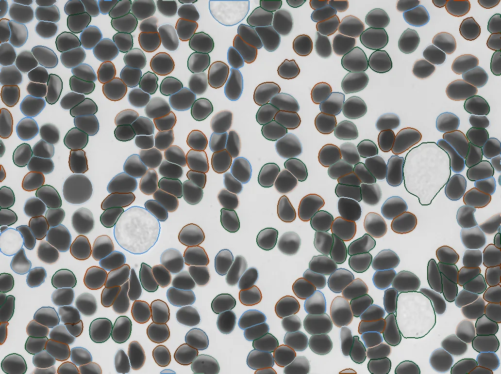
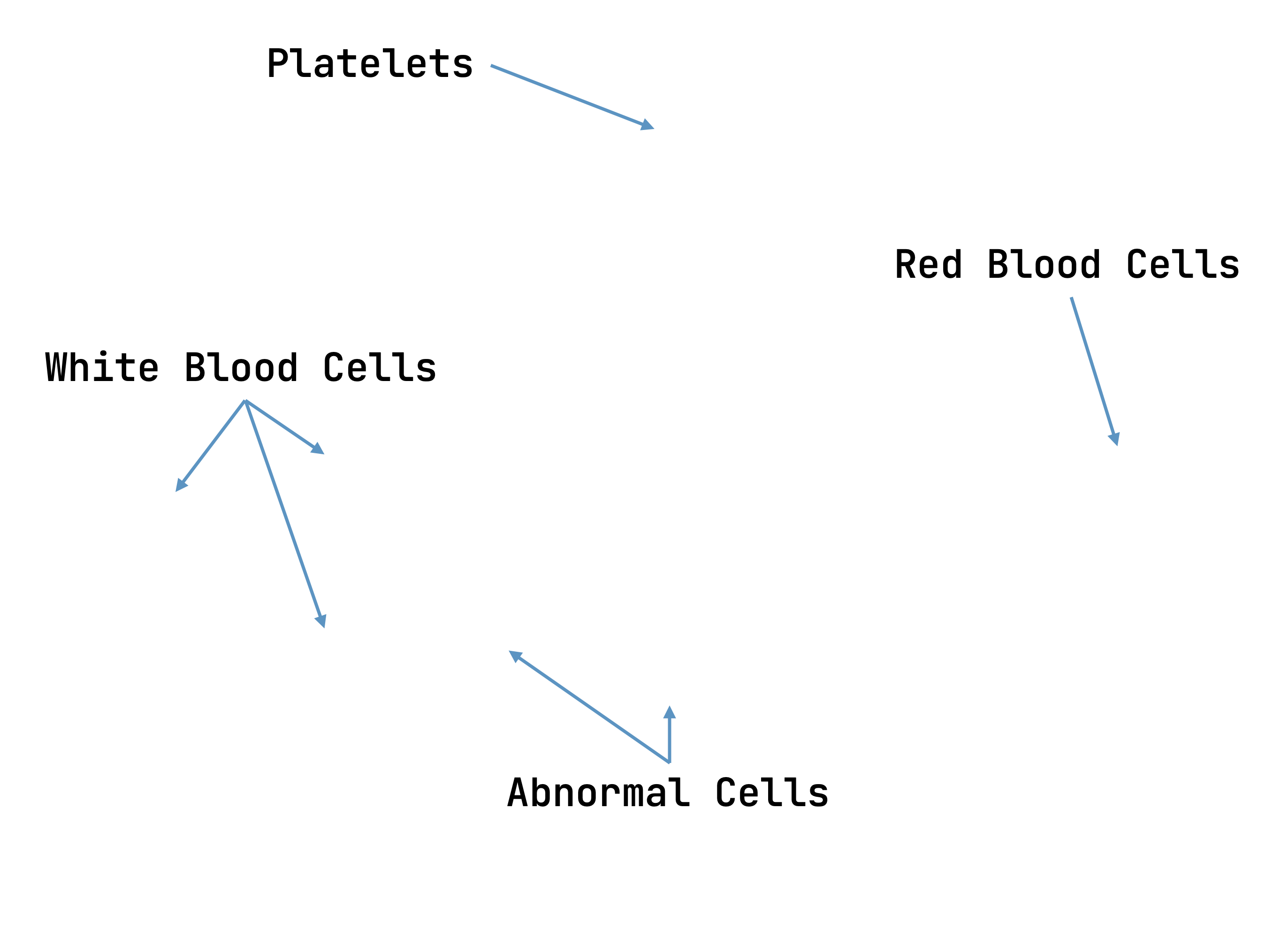 Analysis Mapped by morphology Foundation-model embeddings, fine-tuned per task